Medical imaging and software
EXPERIENCE IN DIFFERENT FIELDS ALLOWS DATAMIND TEAM TO RAPIDLY IDENTIFY THE BEST TECHNOLOGIES AND METODOLOGIES FOR KNOWLEDGE EXTRACTION IN MEDICAL IMAGING.
Medical imaging is the technique and process used to create images of the human body (or parts and function thereof) for clinical purposes (medical procedures seeking to reveal, diagnose, or examine disease) or medical science (including the study of normal anatomy and physiology).
Medical software refers to computer programs designed to assist healthcare professionals in managing patient data, diagnosing illnesses, creating treatment plans, and other medical-related tasks.
DataMind specializes in developing customized software solutions for medical devices. The company works closely with its clients to understand their specific needs and requirements, and develops tailored software components to meet those needs. DataMind also possesses the necessary expertise to work on software that meets the stringent regulatory requirements for medical use. This ensures that the final product is not only functional and tailored to the client's needs, but also meets the high standards necessary for medical software certification.
DataMind has worked in a few projects in this fields, for example:
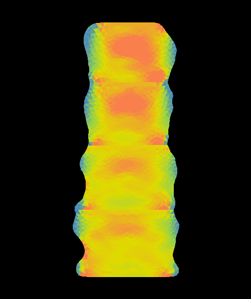
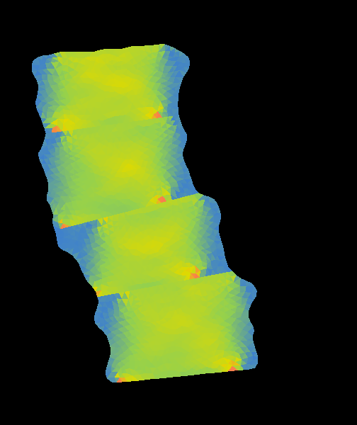
Bone Strain Index
In recent years, the study of bone metabolism has required lot of efforts to consider all the mechanical aspects for a correct assessment of bone strength and associated fracture risk.
Data regarding bone density, structure and geometry are usually obtained by the gold standard method of dual X-ray absorptiometry (DXA).
Despite this, several studies were successful in showing that Finite Element Model strength predictions outperform bone mineral denisity as a predictor for fracture.
In order to automate a Finite Element Model applied to lumbar and femur DXA images, Bone Strain Index Software has been developed in collaboration with Tecnologie Avanzate.
Bone Strain Index Software is regulated by MDR and has been created according to the regulations that define the set of processes, activities, and tasks required to medical device software life cycle.
This project has been realized in collaboration with Tecnologie Avanzate Srl.
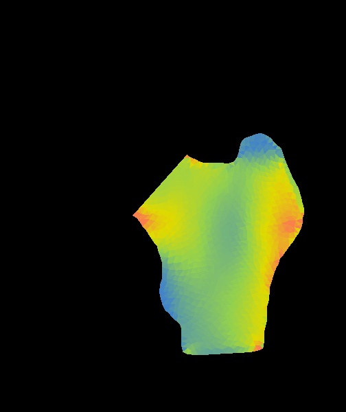
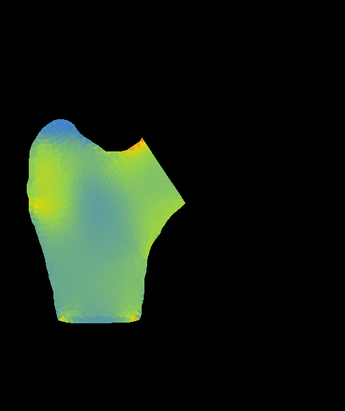
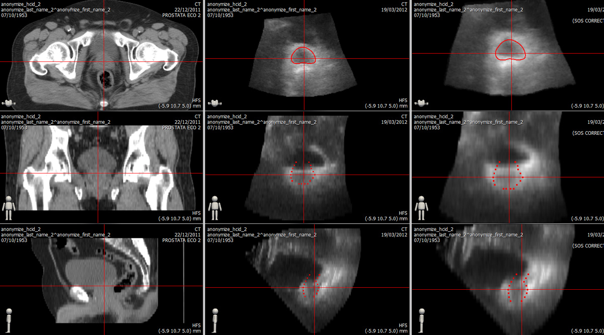
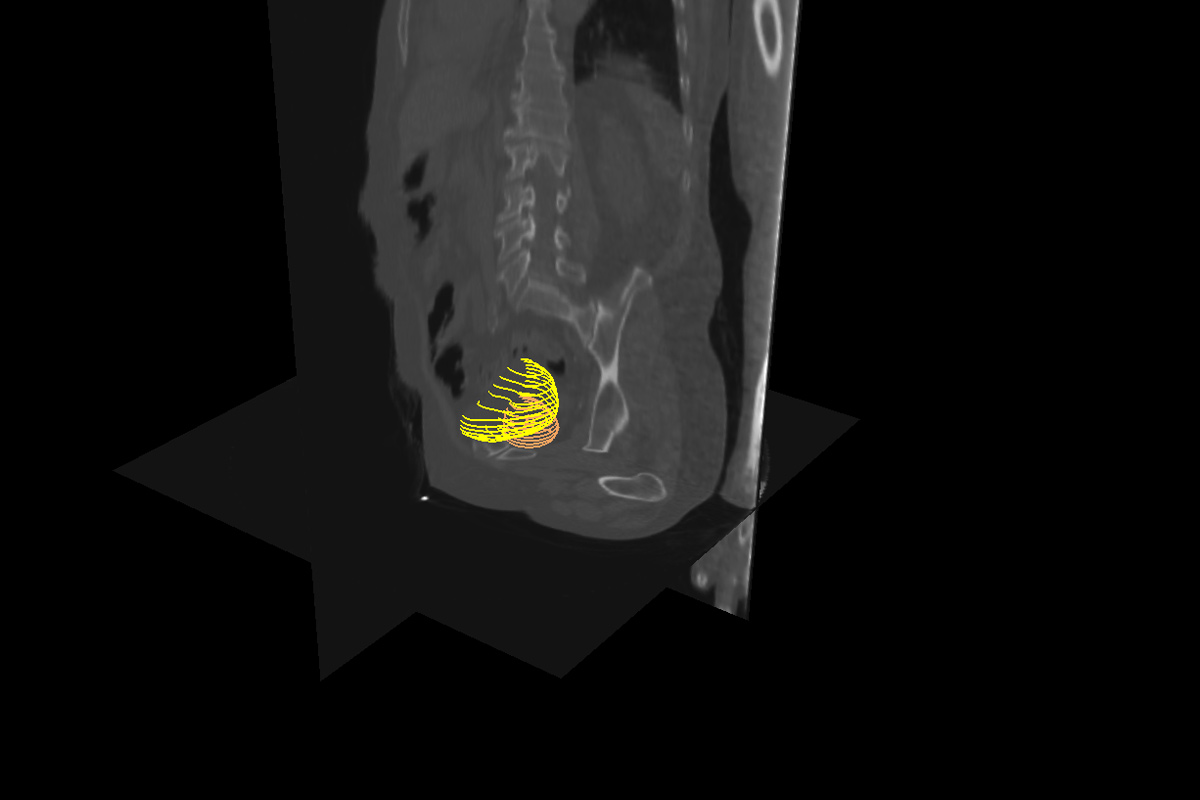
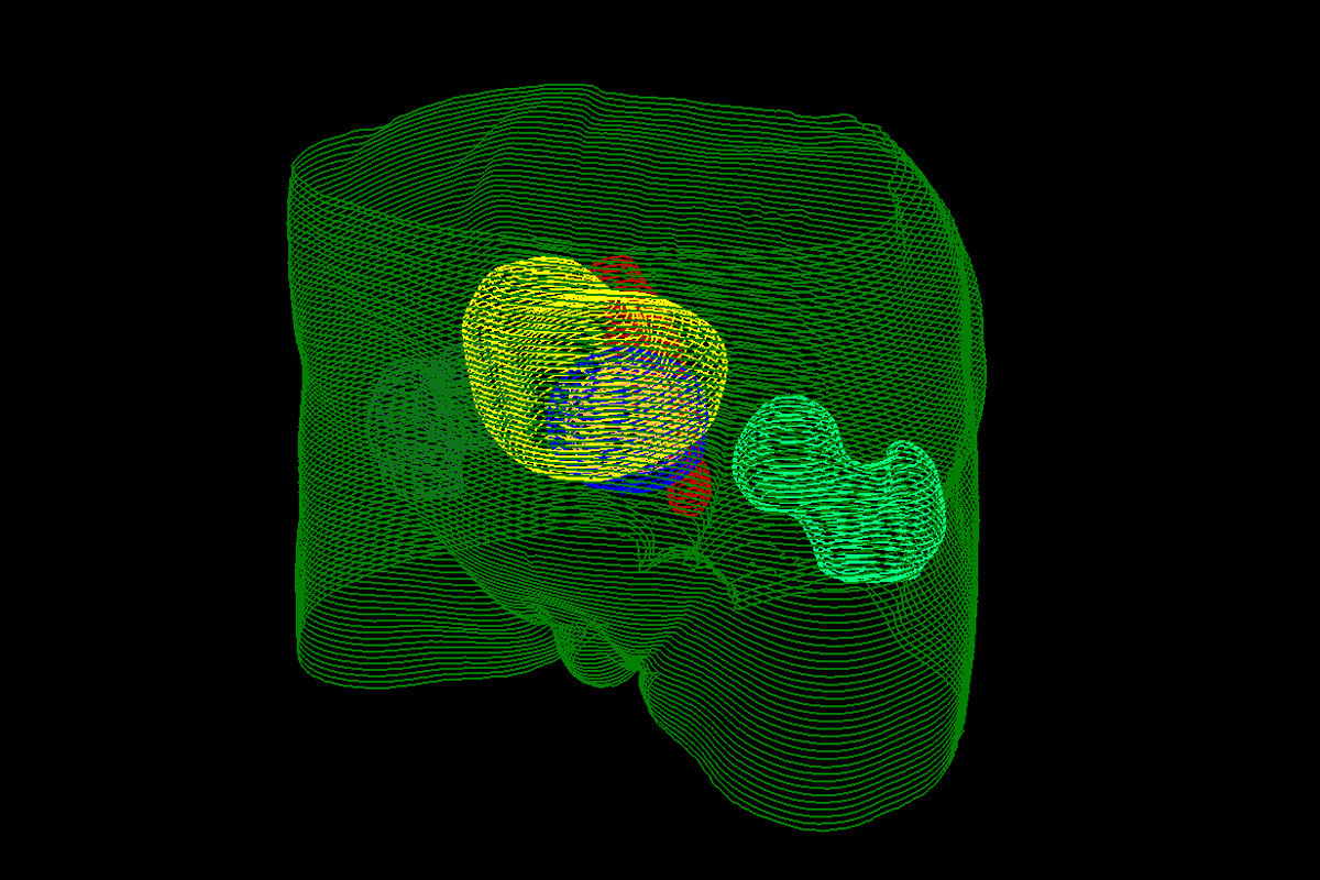
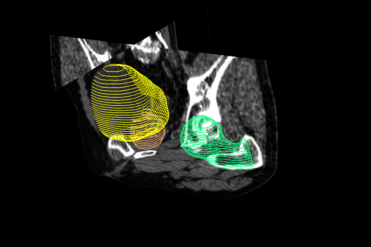
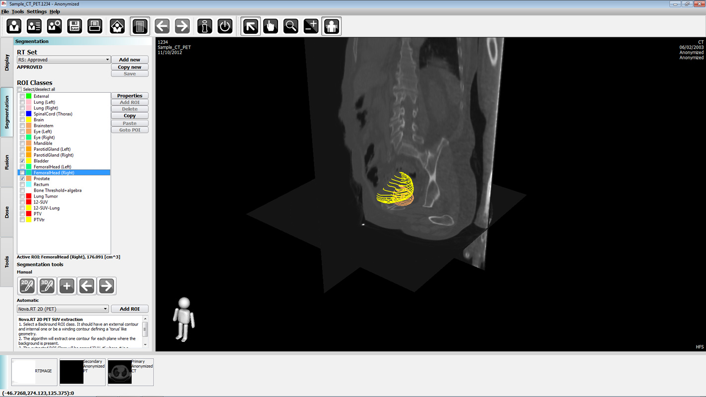
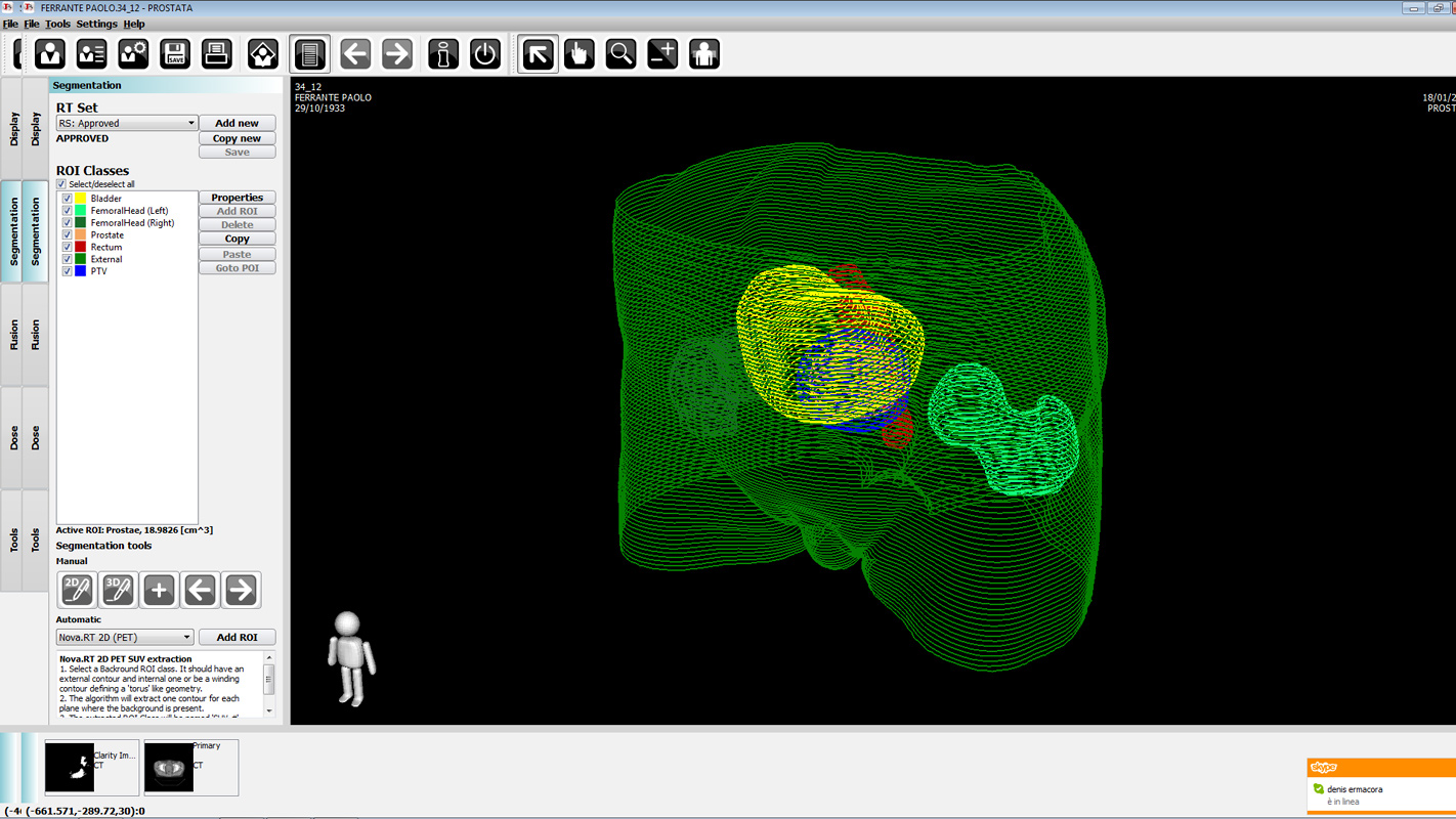
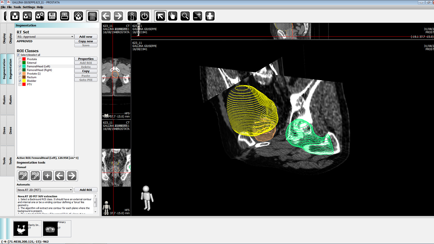
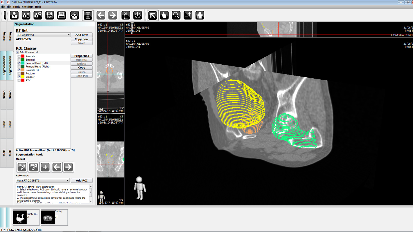
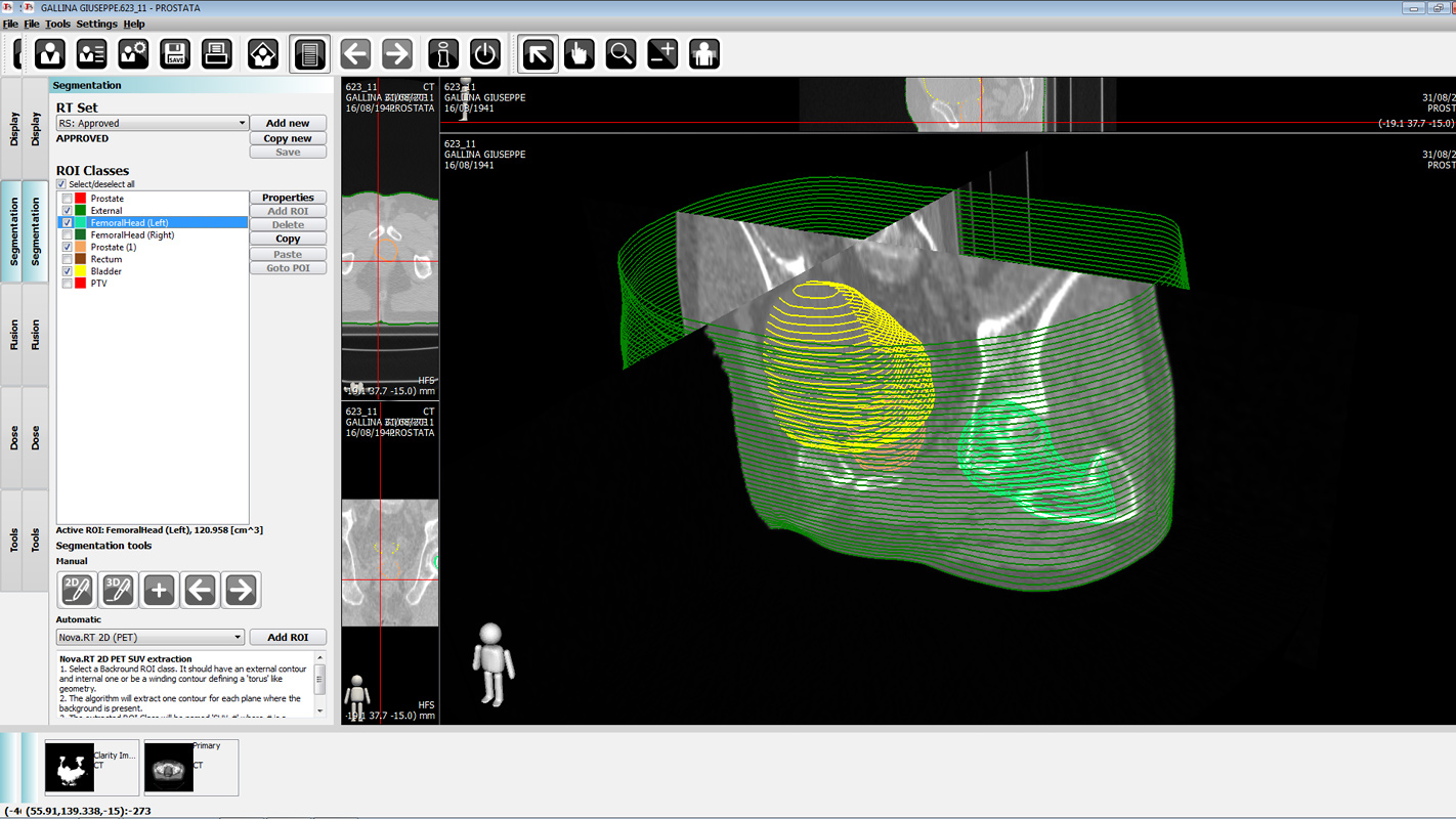
Multi-modality Organ Segmentation - Ultrasound (US) Tomography (CT)
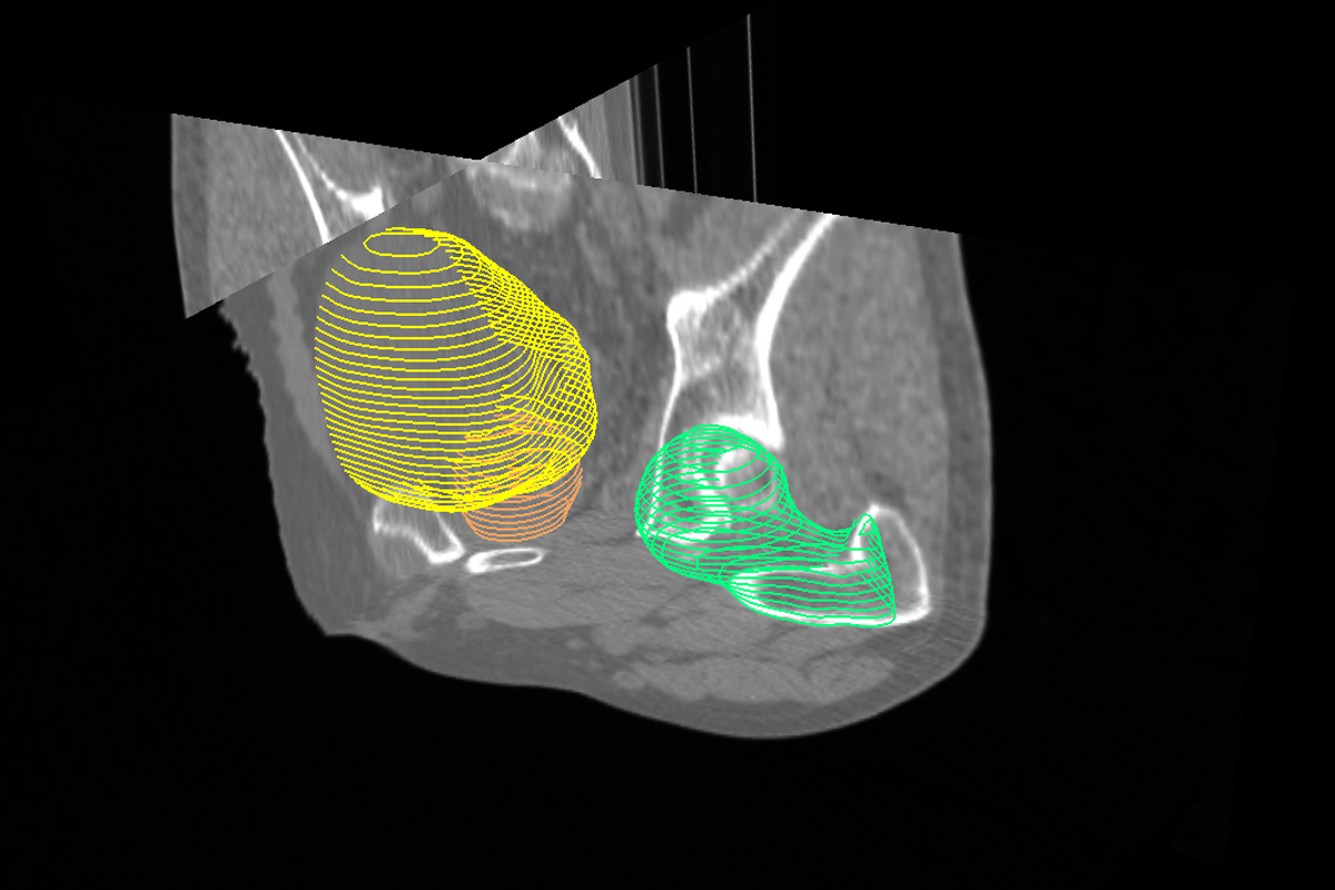
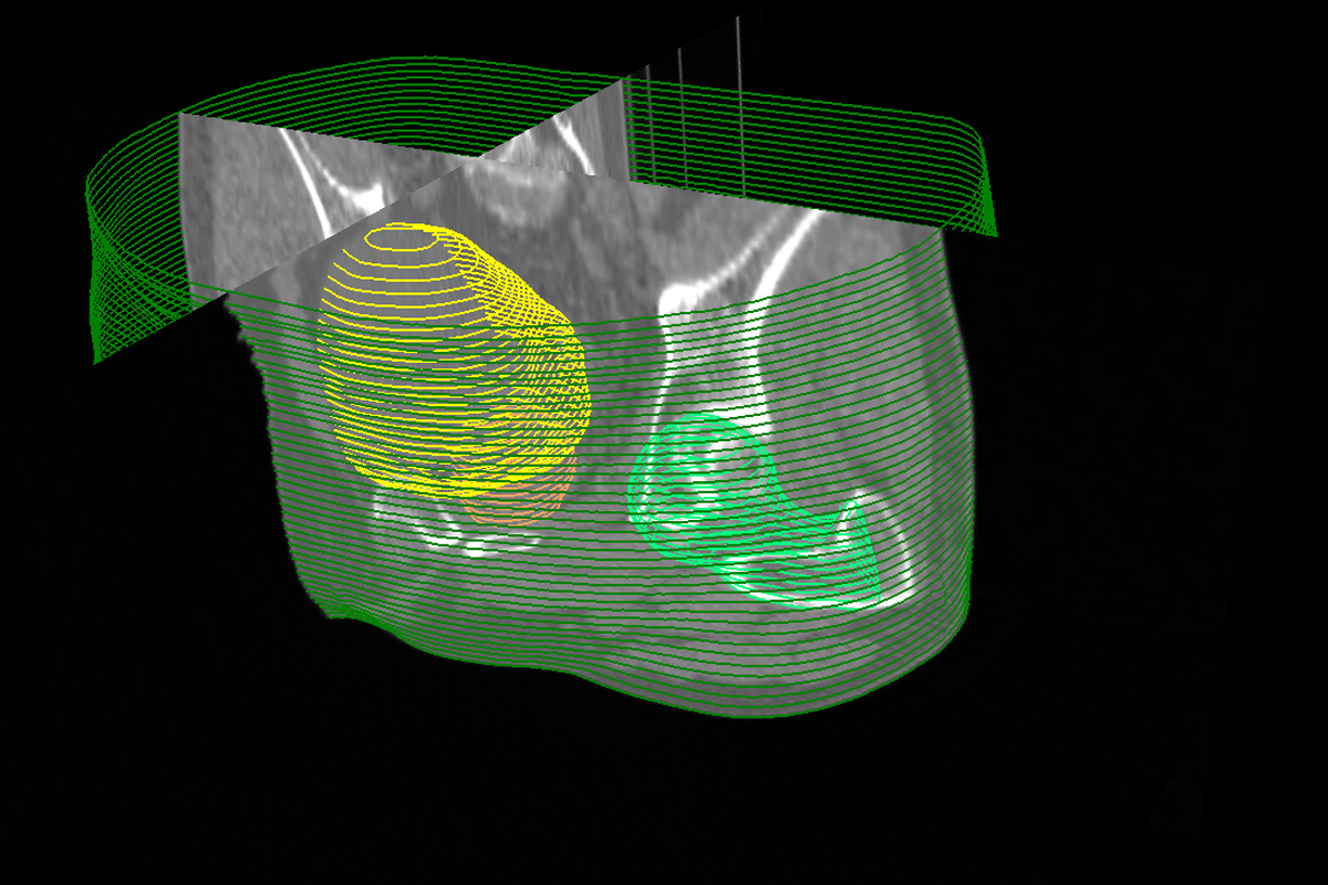
Segmentation of targets and organs at risk in patient images used in radiotherapy is a very delicate task as errors in this phase propagate systematically through the whole treatment course. Moreover, it is becoming more and more time demanding since advanced treatment techniques, such as intensity modulated radiation therapy, were introduced. To improve workflow efficiency, semi-automated contouring techniques were implemented in treatment planning systems, based on computed tomography (CT) imaging datasets. But information from this imaging modality is not always sufficient to provide the necessary accuracy, even for manual contouring. This led to the need of using multiple imaging modalities in this process. Until now, no real automated multi-modality segmentation algorithm was available. In this work we introduce a three dimensional (3D) cross-modality automated image segmentation algorithm based on CT and ultrasound (US) images. The algorithm can be trained and optimized on the characteristics of specific patient samples, becoming increasingly accurate with the size of the sample. We cross-trained and cross-tested the algorithm on sixteen prostate patients. The conformity between the automatically segmented prostate contours and the contours manually outlined by an expert physician on the CT-US fusion was assessed using the Mean Distance to Conformity. In all of these cases the addition of US information reduced the MDC index. The fully automated cross-modality algorithm provided therefore contours that always matched the manually outlined references.
This project has been realized in collaboration with Tecnologie Avanzate Srl.
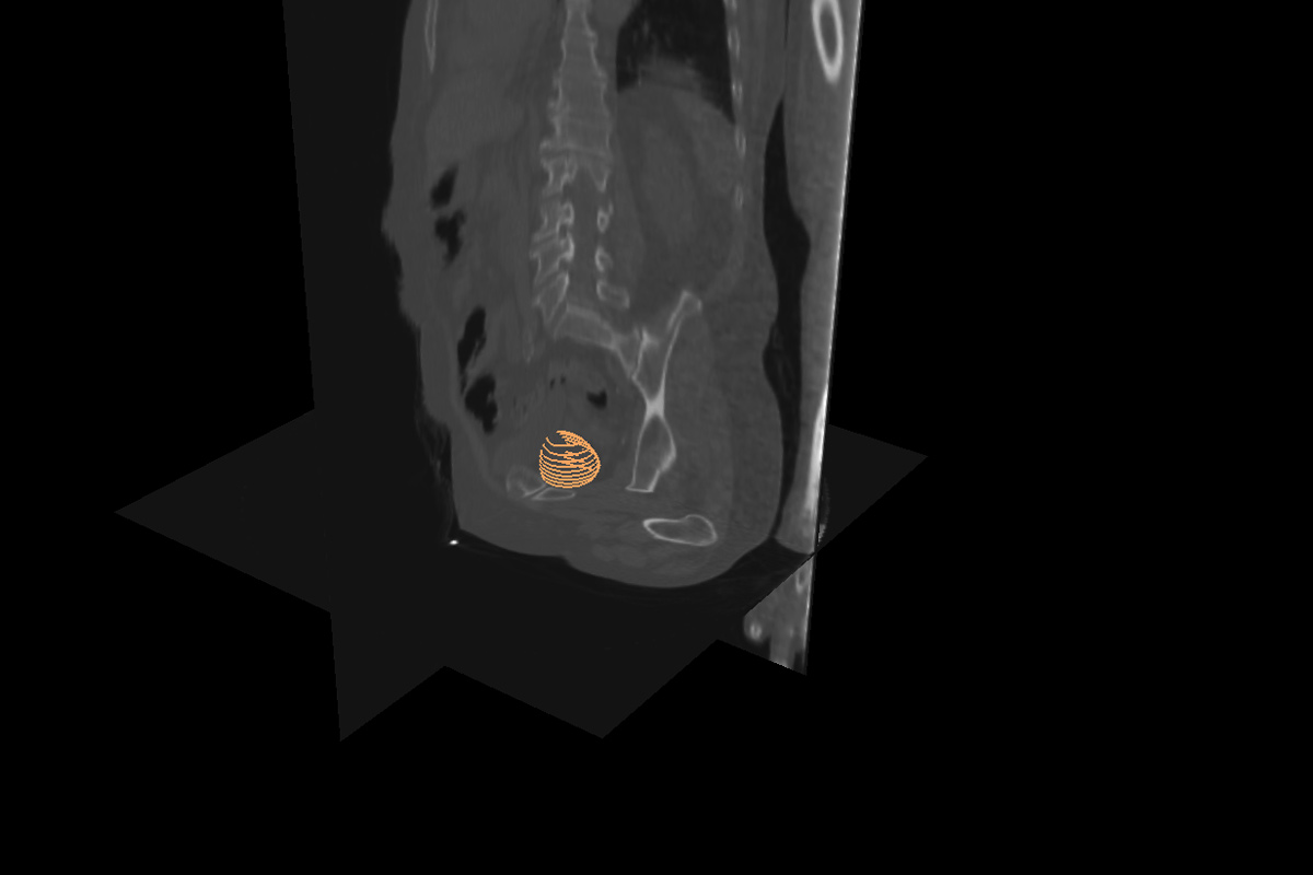
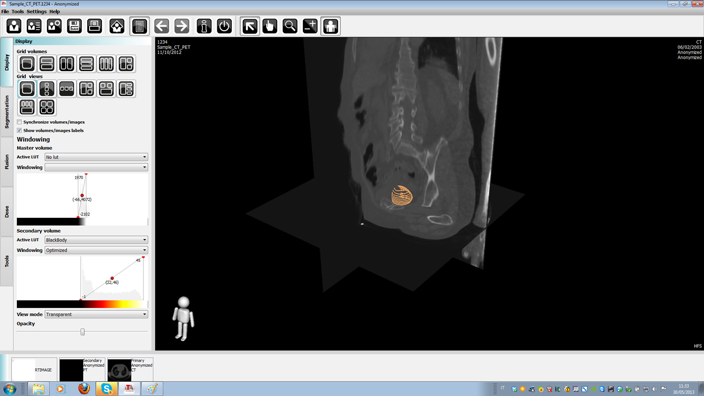
Lesion Detection - Tumor Delineation on PET
Radiation therapy is a local treatment, whose goal is to kill all clonogenic tumor cells and to reach a complete local tumor remission. Traditionally, treatment planning, monitoring and evaluation of response after radiotherapy are mainly based on computed tomography and magnetic resonance. However, concerning target volume delineation for radiotherapy, anatomical imaging has limits in the presentation of tumor extension, when tumor and normal tissues have similar density, similar magnetic properties or a similar contrast enhancement. Therefore, the new imaging methods of biological imaging such as 18F- fluorodeoxyglucose (FDG)-PET/CT imaging are increasingly used to define target volume in radiation treatment planning, although a standardized way of converting PET signals into target volumes is not yet available. To address these issue we developed an automated algorithm that delineates the target volumes determining dynamically the 3D region that minimizes a cost function extrapolated from the results of an important multicentric study in which the major Italian clinics were involved. The algorithm can handle volumes coming from all PET scanners and all kind of not-moving lesions.
This project has been realized in collaboration with Dott. Marco Brambilla, Secretary General of EFOMP, European Federation of Organisations for Medical Physics, Direttore SC di Fisica Sanitaria, Az. Ospedaliero Universitaria Maggiore della Carità di Novara and with Tecnologie Avanzate Srl.